![]() Figure 2 of
Carlson, Mol Vis 2003;
9:615-623.
Figure 2 of
Carlson, Mol Vis 2003;
9:615-623.
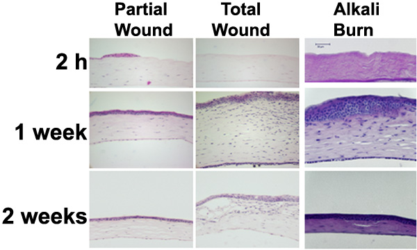
Figure 2. Hematoxylin and eosin staining of mouse cornea sections
Paraffin sections of mouse eyes at 2 h, 1 week, and 2 weeks post-wound stained with hematoxylin and eosin followed by light microscopy. Epithelial removal can be seen at 2 h post-injury in all wound types. Edema is most noticeable at 1 week post-injury especially in the total epithelial debridement and alkali burn. Partial debridement corneas return to a normal morphology 2 weeks post-wound; whereas total epithelial debridement corneas still possess a large amount of edema and conjunctival infiltration.