![]() Figure 5 of
Kanagavalli, Mol Vis 2003;
9:606-614.
Figure 5 of
Kanagavalli, Mol Vis 2003;
9:606-614.
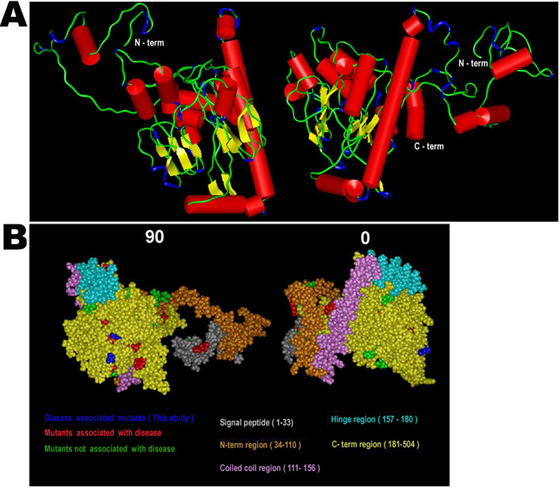
Figure 5. A possible structural model for myocilin
A: The secondary structural elements in the model are colored as follows; Helix (Red), β-strand (Yellow), Turns (Blue) and random coil (green). The N-term and C-term are marked. The N-term region has low secondary structure content and the C-term region is more compact. The long helix in front of the C-term region is the coiled coil region. B: Two orthogonal views of the CPK space-filling model are shown. The view at 90 is equivalent to viewing the model shown in A from behind the page. The color scheme is indicated in the figure.