![]() Figure 1 of
Kanagavalli, Mol Vis 2003;
9:606-614.
Figure 1 of
Kanagavalli, Mol Vis 2003;
9:606-614.
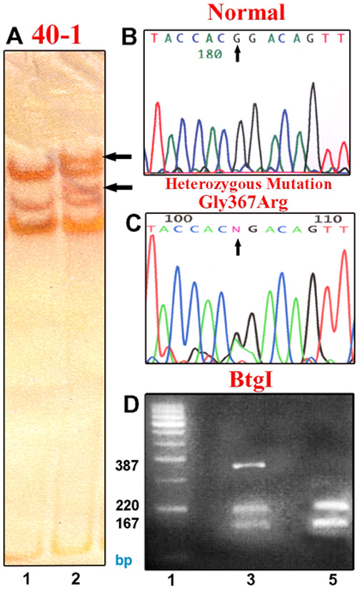
Figure 1. Heterozygous Gly367Arg mutation in exon 3 of MYOC
A: SSCP analysis. Lane 1 is normal control, Lane 2 shows the mobility shift in proband 40-1 as indicated by arrow. B: Forward chromatogram sequence derived from a normal individual where the normal base G at nucleotide 1099 is indicated by arrow. C: Forward chromatogram sequence derived from proband 40-1 showing heterozygous mutation Gly367Arg1099G>A caused an amino acid change from Glycine to Arginine at codon 367. D: Btg I restriction to reconfirm the heterozygous Gly367Arg mutation. Lane 1 is the 100 bp ladder, Lane 3 is proband 40-1 and Lane 5 is an unaffected control.