![]() Figure 2 of
Hartwick, Mol Vis 2003;
9:594-600.
Figure 2 of
Hartwick, Mol Vis 2003;
9:594-600.
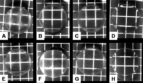
Figure 2. Appearance of cultured lenses recovering from anesthetic-induced optical changes
Photographs of a representative lens 1 h (A, B, C, and D) and 8 h (E, F, G, and H) following a 2 h exposure to Alcaine® (A, E), Fluoracaine® (B, F), or Fluress® (C, G). Control lenses (D, H) are included for comparison. The squares on the grid are 5x5 mm.