![]() Figure 3 of
Zhang, Mol Vis 2003;
9:584-593.
Figure 3 of
Zhang, Mol Vis 2003;
9:584-593.
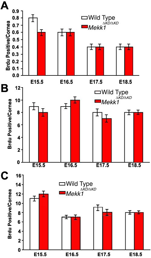
Figure 3. MEKK1 does not affect corneal cell proliferation
Pregnant females of a Mekk1+/ΔKD heterozygote F1 cross were injected with 200 mg/kg BrdU at different gestation days and fetuses from stages E15.5-E18.5 were genotyped and studied for BrdU incorporation. Eye tissues were subjected to immunohistochemistry using anti-BrdU antibodies and counterstained with hematoxylin. The BrdU positive cells were counted in the developing corneal epithelium (A), stroma (B), and endothelium (C). The results represent the average of four independent experiments. The data are expressed as mean±SEM.
