![]() Figure 2 of
Zhang, Mol Vis 2003;
9:584-593.
Figure 2 of
Zhang, Mol Vis 2003;
9:584-593.
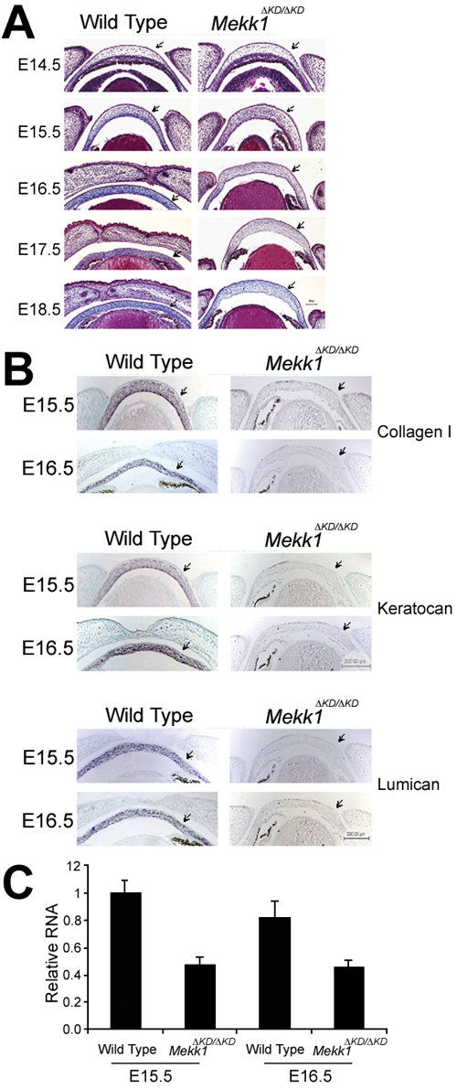
Figure 2. Extracellular matrices deposition in the developing cornea
A: Coronal sections of paraffin-embedded eye tissue of wild type and Mekk1ΔKD/ΔKD fetuses at various days of gestation from E14.5-E18.5 were analyzed by Masson's trichrome staining. B: Wild type and Mekk1ΔKD/ΔKD fetuses at stages E15.5 and E16.5 were analyzed by in situ hybridyzation for mRNAs of Keratocan, Lumican, and Collagen I. Although not obvious at E14.5, the mutant fetuses at E15.5-E18.5 display a reduction in the intensity of staining by Masson's trichrome in the cornea stroma (arrows), wherein the transcripts for Keratocan, Lumican, and Collagen I were also reduced. C: Total RNA isolated from E15.5 and E16.5 fetuses of wild type and Mekk1ΔKD/ΔKD corneal tissue was examined by real-time RT-PCR for Collagen I transcripts. The results represent the average of six experiments with enough RNA needed to reach an arbitrary amplification threshold value normalized to Gapdh mRNA in one sample relative to that in the E15.5 wild type cornea.