![]() Figure 6 of
Davies, Mol Vis 2003;
9:569-578.
Figure 6 of
Davies, Mol Vis 2003;
9:569-578.
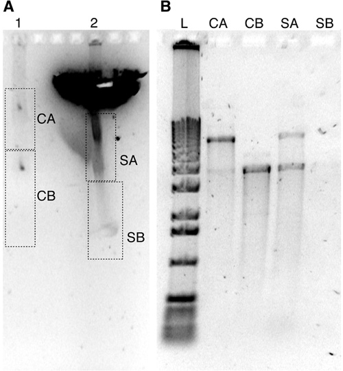
Figure 6. Electrophoresis of plasmid DNA through human sclera
A: Plasmid DNA (pEGFP-1) after standard electrophoresis through 1.1% agarose (lane 1) is compared to that which is impeded by a piece of human sclera (lane 2). The gel was loaded with 925 ng of pEGFP-1 in lanes 1 and 2, subjected to 56 Volts for 2.5 h in 1X TAE, and was stained with SYBR Green for 35 min. Following electrophoresis, displayed in A, DNA was purified from agarose slices taken from each lane (the location of each agarose slice is denoted by the boxes labeled CA, CB, SA, and SB). B: DNA was recovered from each gel slice from A, reduced in volume, loaded on a second 0.8% agarose gel, subjected to 56 V for 1.5 h, and stained with SYBR Green for 30 min. Recovered gel-purified DNA (in B, lane SA) reveals that the smear located below the sclera in A (box SA) contained DNA identical in mobility to plasmid recovered from lanes unimpeded by sclera (lanes CA and CB). Lane SB contained little DNA. These results suggest that the plasmid crossed the sclera. The lane labeled L was loaded with 1,000 ng of a 1 kb ladder (Invitrogen, Carlsbad, CA).