![]() Figure 5 of
Davies, Mol Vis 2003;
9:569-578.
Figure 5 of
Davies, Mol Vis 2003;
9:569-578.
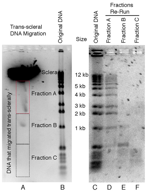
Figure 5. Electrophoresis of linear double stranded DNA fragments through the sclera
Typical results from the electrophoresis of DNA of differing sizes through human sclera. DNA (ranging from approximately 50 bp to 12 kb long) passed through the sclera in the electric field. This experiment was repeated five times; data shown are representative. Lane A shows a DNA ladder (1 kb ladder; Invitrogen Corporation, Carlsbad, CA) that passed through the sclera. Lane B shows the same DNA ladder unimpeded by sclera. The lane A material that had passed through the sclera was retrieved in three fractions labeled Fractions A, B, and C, as illustrated by the dotted lines (the Fraction A box is in red to indicate the upper boundary of the excised gel, which was about 2 mm downstream of the scleral fragment). The DNA from each agarose gel fragment was recovered and analyzed on a second gel. Wells D, E, and F were loaded with DNA extracted from Fractions A, B, and C, respectively. The original DNA ladder is shown in Lane C. There was no scleral tissue in Fraction A. Fraction A (Lane D) shows that DNA as large as 12 kbp migrated through the sclera. The mobility of the smaller double-stranded DNA fragments is largely unaffected. In larger fragments, from about 400 bp and greater, mobility is reduced in proportion to the size of the DNA.