![]() Figure 3 of
Davies, Mol Vis 2003;
9:569-578.
Figure 3 of
Davies, Mol Vis 2003;
9:569-578.
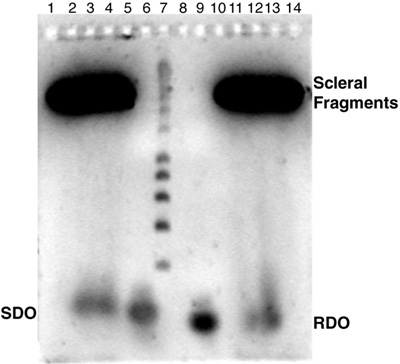
Figure 3. Electrophoresis of small oligonucleotides through scleral fragments
The same experimental apparatus and protocols were used as described in Methods and the legend for Figure 2, except small oligonucleotides were loaded into sample wells in 10% glycerol 1X TAE buffer. Two types of oligonucleotides (SDO and RDO) were loaded in the wells to test whether they could be electrophoresed through the sclera. First, a 51-base long single stranded DNA (SDO) was tested (see lanes 3 and 6). Lane 3 contained a scleral fragment and lane 6 did not. The second type of oligonucleotide was a hybrid RNA-DNA molecule (RDO) possessing a double hairpin structure synthesized as a 68-mer. RDO was loaded in lanes 9 and 12. Lane 12 contained a scleral fragment whereas lane 9 did not. The gel was run at 58 volts for 2 h and 5 min. Subsequently, the gel was soaked in SYBR Green II (which effectively stains both single stranded and double stranded DNA) for 30 min and examined by fluorescence.