![]() Figure 2 of
Davies, Mol Vis 2003;
9:569-578.
Figure 2 of
Davies, Mol Vis 2003;
9:569-578.
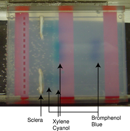
Figure 2. Electrophoresis of charged dyes through human sclera
Rectangular fragments of human cadaveric sclera were mounted vertically in a horizontal gel electrophoresis chamber. The tissue samples were oriented with the outside eye surface facing and closest to the gel wells. 2% agarose was poured into the gel chamber and allowed to solidify, which embedded the sclera and formed wells. The agarose contained 1X TAE buffer (40 mM Tris-acetate, 1 mM EDTA, pH 8.0). Bromphenol blue and xylene cyanol in 1X TAE buffer and 10% glycerol were loaded into well positions 3, 6, 8, and 12. Two samples (Lanes 6 and 8 from the top down) were unimpeded by sclera, and two other wells (lanes 3 and 12) were completely occluded by human scleral fragments. The samples were subjected to an electric field of 3.3 V/cm for 114 min, and digital photographs were taken every 3 min during electrophoresis. The final picture of the series is shown (A). The 39 pictures of the run are shown in sequence as a movie (B). Lanes 6 and 8 show that bromphenol blue migrated roughly twice as quickly (about 4 cm/h) as xylene cyanol (about 2 cm/h) when unimpeded by sclera. Lanes 3 and 12 show that bromphenol blue ran into the sclera first, but its passage was slowed or delayed in this encounter while xylene cyanol met the sclera second. Xylene cyanol in lanes 3 and 12 appeared to pass through the sclera relatively unimpeded except for some band spreading when compared to the movement of xylene cyanol in control lanes 6 and 8 that lacked the scleral obstacle. After the xylene cyanol passed through the sclera, bromphenol blue was observed slowly appearing in agarose on the inner side of the scleral face. The movie (B) illustrates that the dyes were not migrating around the sclera. Bromphenol blue was retained and slowly passed through human sclera while a different dye, xylene cyanol, simultaneously passed through the same scleral fragments with minor band spreading and nearly the same migration rate as xylene cyanol through agarose alone.
| This animation requires Quicktime 6 or later. Quicktime is available as a free download. |