![]() Figure 1 of
Davies, Mol Vis 2003;
9:569-578.
Figure 1 of
Davies, Mol Vis 2003;
9:569-578.
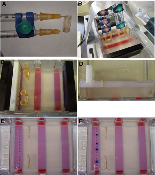
Figure 1. The experimental apparatus
The study design was to conduct experiments with cadaveric human sclera to assure that the same thickness and pore characteristics of sclera would be tested as might be required in treating a human patient. Conventional molecular biological equipment and methods for analyzing DNA were adapted to test whether DNAs of various sizes could be driven across the sclera. The arrangement and assembly of scleral fragments in an agarose gel apparatus is shown in A through F. A: Mounting the scleral fragments on syringe needles before embedding in agarose. B: Securing the mounted scleral fragments during agarose gel solidification. C: Removal of the syringe supports. D: Side view of the embedded scleral fragments. Note the plastic boosts elevating the gel comb. E: Top view of the embedded scleral fragments. F: Loaded DNA and dyes prior to electrophoresis.