![]() Figure 9 of
Al-Shabrawey, Mol Vis 2003;
9:549-558.
Figure 9 of
Al-Shabrawey, Mol Vis 2003;
9:549-558.
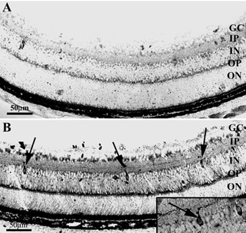
Figure 9. NADPH-diaphorase labeling of frozen retinal sections
At P9, the distribution of NADPH-diaphase reactivity is similar in eNOS+/+ (A) and eNOS-/- (B) retinas, but labeled amacrine-like cells (arrows) are more abundant in the eNOS-/- retinas. Note the prominent axonal labeling of the amacrine-like cells (inset in B) and that the NADPH-diaphorase reaction is stronger in the ganglion cell and the inner and outer plexiform layers of the eNOS-/- retina.