![]() Figure 5 of
Al-Shabrawey, Mol Vis 2003;
9:549-558.
Figure 5 of
Al-Shabrawey, Mol Vis 2003;
9:549-558.
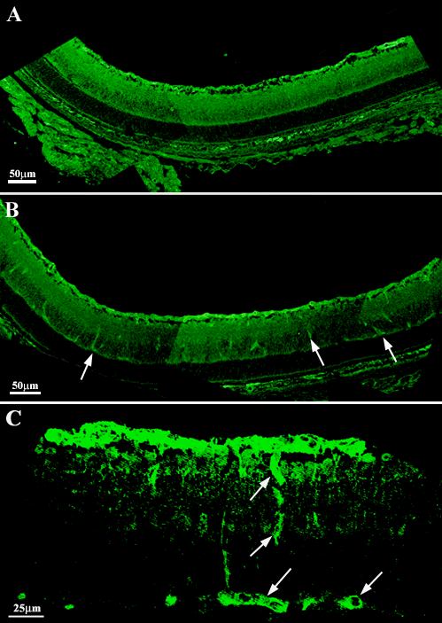
Figure 5. nNOS immunoreactivity in retinal sections from eNOS+/+ and eNOS-/- mice
nNOS immunoreactivity in eNOS+/+ (A) and eNOS-/- (B and C) retinas at P9. nNOS immunoreactivity is present within the nerve fiber, ganglion cell, inner nuclear and inner and outer plexiform layers of both strains. Perivascular labeling is largely restricted to the superficial vessels in the eNOS+/+ retinas, whereas the deeper vascular layers are strongly immunoreactive in the eNOS-/- retinas (arrows in B and C).