![]() Figure 4 of
Al-Shabrawey, Mol Vis 2003;
9:549-558.
Figure 4 of
Al-Shabrawey, Mol Vis 2003;
9:549-558.
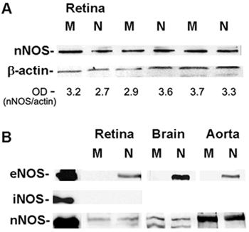
Figure 4. Western blotting analysis of nNOS, eNOS, and iNOS protein levels
Protein samples (50 μg from 2 retinas/lane) prepared from eNOS-/- mice (M) at P9 (A) show nNOS levels similar to those in age-matched eNOS +/+ mice (N). Immunoblots were re-probed with an antibody against β-actin to control for possible differences in protein loading. Densitometric analysis showed no significant difference in relative optical density between the two groups (p>0.05, unpaired t-test, n=3). Immunoblot analysis of tissue extracts (50 μg protein) prepared from pooled retinas (n=10), abdominal aortas (n=3) and brain capillaries (n=3) of P30 mice (B) demonstrates that eNOS protein is present in eNOS+/+ mice (M), but absent in the eNOS-/- mice (N). nNOS protein is detected at similar levels in retina, brain capillaries and aorta in both strains. Retina extracts are negative for iNOS protein. The first lane in each blot contains positive control proteins (eNOS=135 kD, nNOS=160 kD, iNOS=130 kD).