![]() Figure 2 of
Al-Shabrawey, Mol Vis 2003;
9:549-558.
Figure 2 of
Al-Shabrawey, Mol Vis 2003;
9:549-558.
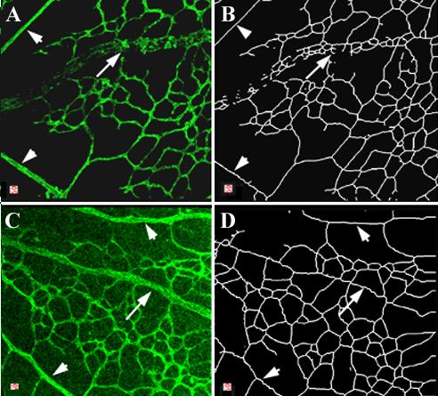
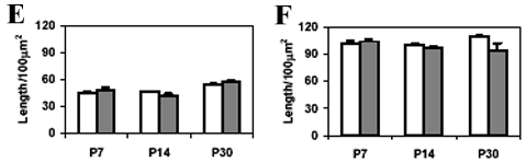
Figure 2. Lectin labeling of retinal flat mounts from eNOS+/+ and eNOS -/- mice
Lectin-labeled whole-mount retinas from eNOS+/+ (A, B) and eNOS-/- (C, D) mice. At P7 the radial pattern of retinal vessels is established. The arteries (arrowheads) are surrounded by capillary free zones, while the capillary networks around the veins (arrows) are continuous. For analysis of capillary density, the vessels located between the major arteries and veins were reduced digitally to 1 pixel in diameter (B, D) and their length/100 μm2 was determined by computer-assisted morphometry. This analysis showed no significant differences between the eNOS+/+ (open bars) and eNOS-/- (filled bars) mice in either the central (E) or peripheral (F) portions of the retina (p>0.05, ANOVA, n=5 mice per group, error bars represent the standard error of the mean).