![]() Figure 1 of
Al-Shabrawey, Mol Vis 2003;
9:549-558.
Figure 1 of
Al-Shabrawey, Mol Vis 2003;
9:549-558.
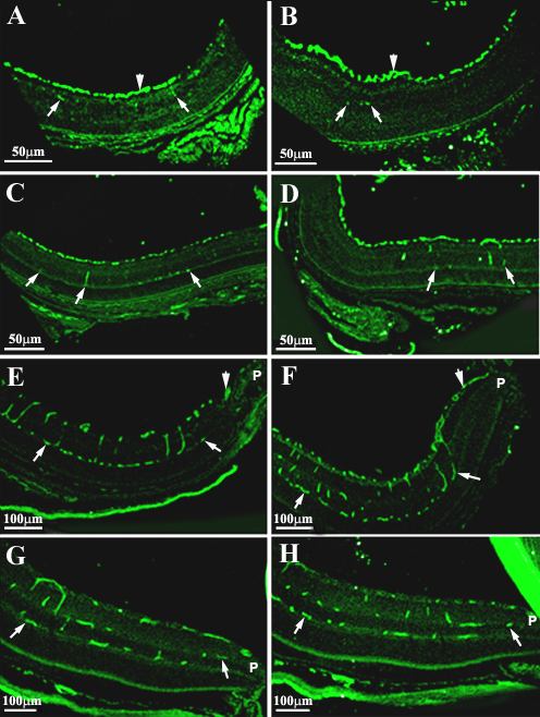
Figure 1. Lectin labeling of retinal frozen sections from eNOS+/+ and eNOS-/- mice
Lectin-labeled frozen retinal sections from eNOS+/+ (A, C, E, G) and eNOS-/- (B, D, F, H) mice. At P7 (A, B) the superficial vascular bed is clearly evident (arrowheads) and a few penetrating vessels have begun to form (arrows). At P9 (C, D) the developing deep vessels extend towards the inner plexiform layer. At P14 (E, F) the superficial vessels (arrowheads) reach the periphery (P), and the deeper vessels (arrows) extend to both inner and outer plexiform layers with communicating channels between. At P30 (G, H) the deep vascular plexus reaches the periphery (P).