![]() Figure 1 of
Huang, Mol Vis 2003;
9:502-507.
Figure 1 of
Huang, Mol Vis 2003;
9:502-507.
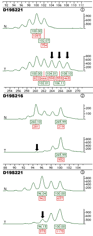
Figure 1. Electropherogram showing examples of LOH and MSI
For each microsatellite marker, the lower trace shows the tumor (T) sample, and the upper trace shows the corresponding normal (N) peripheral blood sample. The X-axis shows size in basepairs (green color), and the Y-axis shows peak height in fluorescence units (red color). Black arrows point to alleles demonstrating microsatellite instability (MSI) or loss of heterozygosity (LOH): MSI at D19S221, LOH at D19S216 (complete loss of smaller allele) and D19S221 (partial loss of smaller allele, AIF=0.211, p<0.5).