![]() Figure 3 of
Proulx, Mol Vis 2003;
9:473-481.
Figure 3 of
Proulx, Mol Vis 2003;
9:473-481.
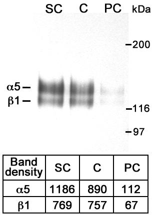
Figure 3. Immunoprecipitation of biotinylated cell surface proteins with anti integrin α5 antibody
Upper panel. Sub-confluent (sc), confluent (c) and post-confluent (pc) primary human RPE cells were harvested, labeled with soluble sulfo-NHS-LC-Biotin and immunoprecipitated with anti-integrin α5 antibody under denaturing conditions. Lower panel. Band density was calculated by the QuantityOne Image analysis software (see Methods).