![]() Figure 1 of
Proulx, Mol Vis 2003;
9:473-481.
Figure 1 of
Proulx, Mol Vis 2003;
9:473-481.
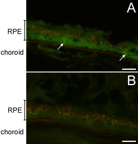
Figure 1. Immunofluorescent detection of integrin α5 in situ
Figure shows cryosections of a retina of a 21 year old donor. As described in methods, specific immunoreactivity appears green and lipofuscin autofluorescence appears orange/yellow. A: Integrin α5 subunit is detected diffusely throughout the cell surface, and more intense staining is found at the basal surface of the RPE monolayer (arrows). The retina peeled off during cryosection processing. B: Negative control using normal rabbit serum. Scale bar represents 10 μm.