![]() Figure 8 of
Fei, Mol Vis 2003;
9:31-42.
Figure 8 of
Fei, Mol Vis 2003;
9:31-42.
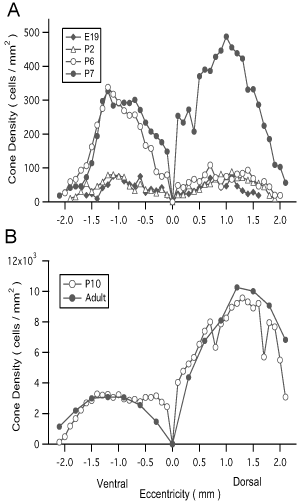
Figure 8. Fluorescent cone densities in the developing and mature retinas
The average numbers of cones expressing the GFP per mm2 retinal area along the dorsal to ventral meridian through the optic disc (OD) were counted from montage images of the retinal wholemounts, and plotted against the retinal eccentricity (mm from the edge of the OD). Three retinas at each time point were sampled and analyzed.