![]() Figure 6 of
Fei, Mol Vis 2003;
9:31-42.
Figure 6 of
Fei, Mol Vis 2003;
9:31-42.
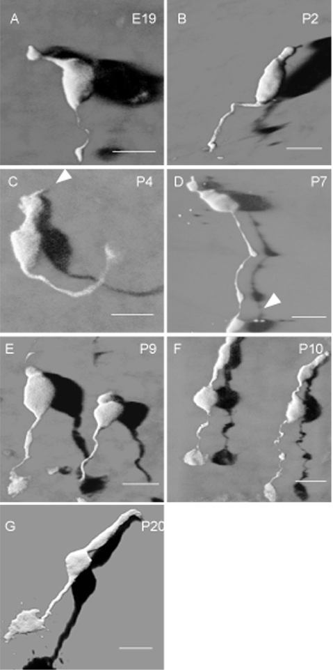
Figure 6. Morphological development of the fluorescent cone cells
These are 3D confocal images of the representative developing fluorescent cells taken from retinal wholemounts of the transgenic mice aged at E19, P2, P4, P7, P9, P10 and P20. Apparent outer segments begin to develop on P4 (C, arrowhead). The cone pedicles with fine basal processes appear in some cones by P7 (D, arrowhead). The cone cells are fully mature by P20 (G). The shadows in these images were generated by shadow projections of the corresponding confocal Z-stack images for 3D view of the cells. Scale bar represents 10 μm.