![]() Figure 5 of
Fei, Mol Vis 2003;
9:31-42.
Figure 5 of
Fei, Mol Vis 2003;
9:31-42.
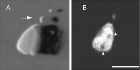
Figure 5. Common morphological features of the E15 fluorescent cells in different mouse lines
These are confocal images of a representative fluorescent cell in the live E15 retina from another transgenic line 5922. A: 3D image, showing the cell body and a sclerally oriented short process (arrow). B: Optical sectioning through the cell body, revealing that the cell had a typical large, elongated nucleus containing 2-3 darker areas (arrowheads) that appear to represent the characteristic dense heterochromatin clumps of the mouse cone nuclei. Scale bar represents 10 μm.