![]() Figure 4 of
Fei, Mol Vis 2003;
9:31-42.
Figure 4 of
Fei, Mol Vis 2003;
9:31-42.
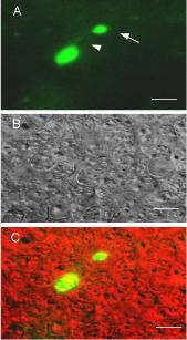
Figure 4. Fluorescent cells are identifiable on E15
Images were collected from the dorsal area of a live, unfixed retina of an E15 transgenic mouse from line 5933, with the microscope focusing on the outer surface of the neuroblast layer. A: Pseudocolored (green) epifluorescence microscope image taken with FITC optics, showing a fluorescent cell bearing a process (arrowhead) with bulbous terminal enlargement (arrow). B: DIC image, showing the profiles of the surrounding non-fluorescent retinal cells of the same field. C: The merged pseudocolor image. Scale bar represents 10 μm.