![]() Figure 3 of
Fei, Mol Vis 2003;
9:31-42.
Figure 3 of
Fei, Mol Vis 2003;
9:31-42.
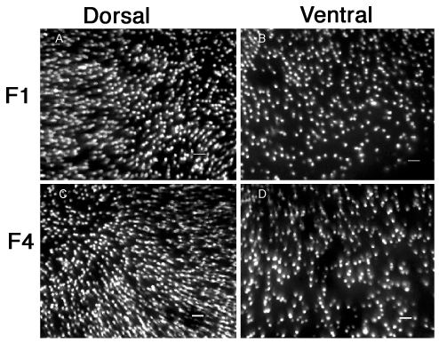
Figure 3. Consistent pattern of GFP expression and the mosaic organization of mouse cones
These are images of fluorescent cones in the dorsal (A and C) and the ventral (B and D) areas of the retinal wholemounts from one first generation (F1; A and B) and one fourth generation (F4; C and D) adult transgenic mouse. They were taken with an epifluorescence microscope with a 25x lens focusing on the cone cell bodies, showing the consistent dorsal-ventral graded GFP expression pattern across mouse generations and the appreciable mosaic organization of the mouse cones. Scale bar represents 20 μm.