![]() Figure 2 of
Fei, Mol Vis 2003;
9:31-42.
Figure 2 of
Fei, Mol Vis 2003;
9:31-42.
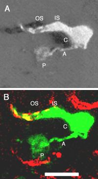
Figure 2. Fluorescent photoreceptor cells are cones
These are confocal images of a representative fluorescent cell taken from a PNA labeled flat mounted retina. A: 3D image showing the morphological features of mouse cones including a conical shaped outer segment (OS), a thick inner segment (IS) and a large synaptic pedicle (P) connected to the cell body (C) via an axon (A). B: Co-labeling of this fluorescent cone (green) by cone specific PNA marker (red) in the outer segment (OS). Note that these images were from the mid-peripheral ventral retina. The cone cell does not appear to stand upright, which is likely caused by the coverslipping process that might push the cells to bow forward. There are 2-3 PNA+ cells in B that did not express GFP, which is common in the ventral retina. Scale bar represents 10 μm.