![]() Figure 11 of
Fei, Mol Vis 2003;
9:31-42.
Figure 11 of
Fei, Mol Vis 2003;
9:31-42.
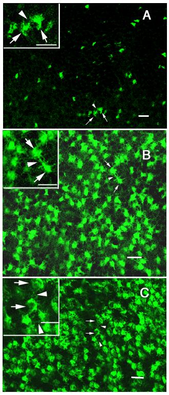
Figure 11. Developmental patterning of cone pedicles in the outer plexiform layer
These projected confocal images of the representative fluorescent cone pedicles were taken from the dorsal retinas of the P9 (A), P10 (B) and adult (C) transgenic mice. The insets were enlarged images to show the fine processes of the cone pedicles. On P9, some fluorescent cones exhibited enlarged axonal terminals, while others developed pedicles with basal processes (A, arrowhead) that contact pedicles (A, arrows) of the neighboring cones. But patterning of the cone pedicles in the OPL was incomplete. The cone pedicles were well developed by P10, with extensive basal processes (B, arrowheads) contacting other cone pedicles (B, arrows). At this stage, the morphology of individual pedicles and the spatial arrays of all pedicles in the OPL were very similar to that in the adult retina (C, arrows: pedicles; arrowheads: basal processes). Scale bar represents 10μm.