![]() Figure 1 of
Fei, Mol Vis 2003;
9:31-42.
Figure 1 of
Fei, Mol Vis 2003;
9:31-42.
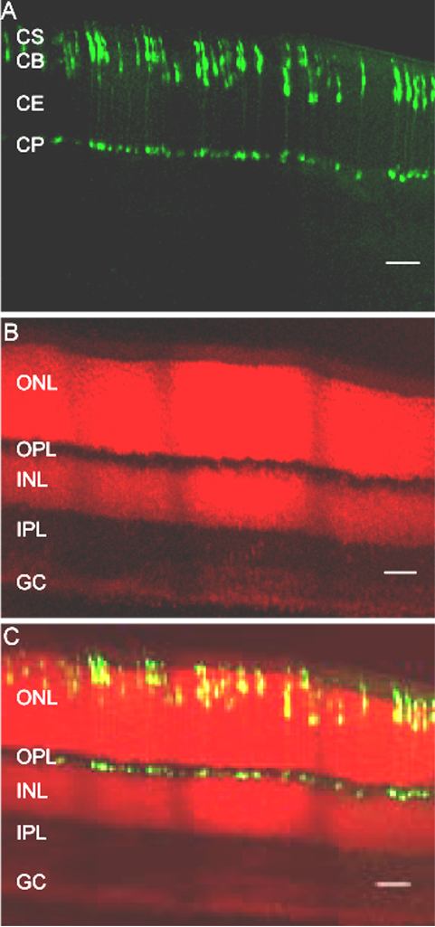
Figure 1. Fluorescent cells are restricted to the photoreceptor cell layer
These are projected confocal images from 36 frames of a propidium iodide counterstained cross retinal section from an adult transgenic mouse taken with standard FITC/Rhodamine filter sets and a 20x lens. A: Image (pseudocolored green) of the GFP expressing fluorescent cells. B: Rhodamine image (pseudocolored red) of the same filed, showing the three nuclear layers and the two synaptic layers of the mouse neural retina. C: The merged image, showing the location of the majority of fluorescent cell bodies in the outer border of the outer nuclear layer (ONL) and the cone pedicles (CP) in the inner part of the outer plexiform layer (OPL). No cells in other retinal layers exhibit GFP fluorescence. CS: outer and inner segments of photoreceptor cells. CB: cell bodies. CE: axons. INL: inner nuclear layer. IPL: inner plexiform layer. GC: ganglion cell layer. Scale bar represents 50 μm.