![]() Figure 4 of
Zhang, Mol Vis 2003;
9:465-472.
Figure 4 of
Zhang, Mol Vis 2003;
9:465-472.
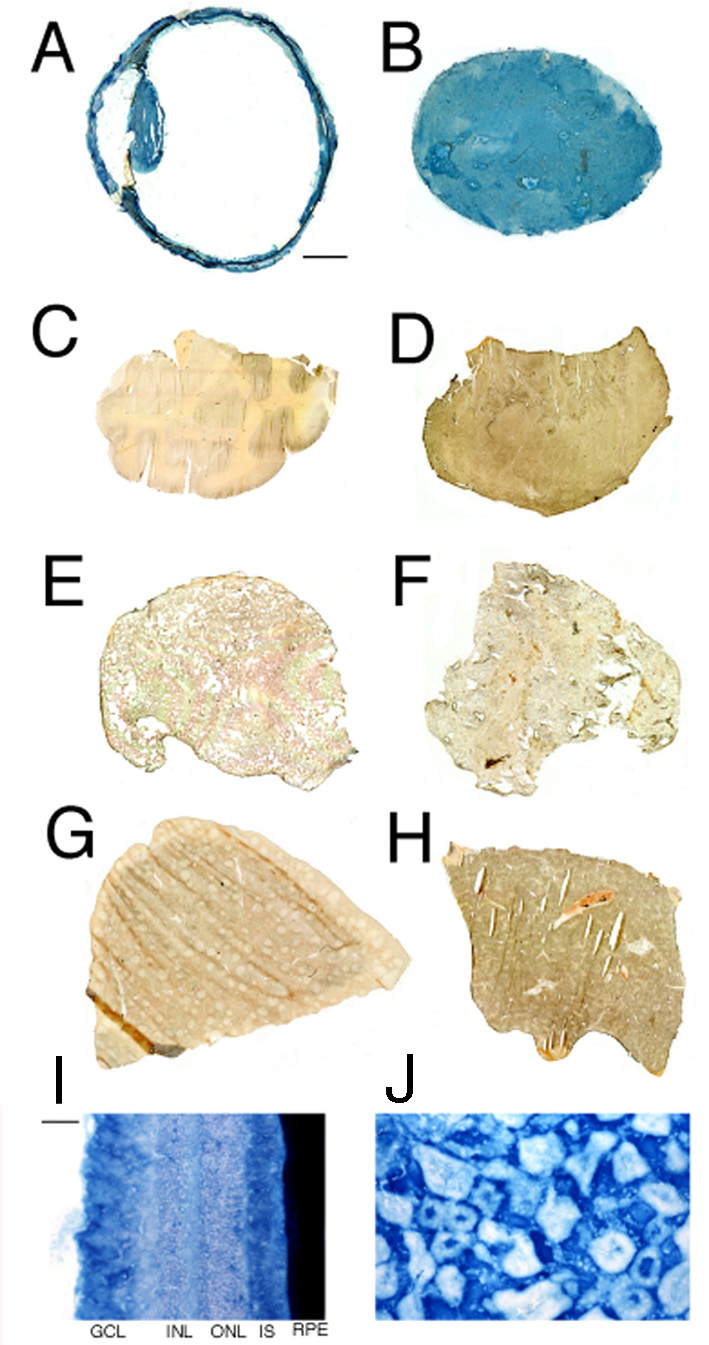
Figure 4. β-Galactosidase histochemistry of primate organs
Organ β-galactosidase histochemistry for Rhesus monkey eye (A and I), kidney (B and J), brain (C), heart (D), lung (E), omental fat (F), spleen (G), and liver (H). Skeletal muscle was also examined (not shown), and like the heart showed no histochemical reaction. The organs were removed from the animal sacrificed at 48 h after an intravenous injection of the opsin promoter-driven pLacF plasmid encapsulated in HIRMAb-targeted PILs, which corresponds to monkey 2 (see Methods). The magnification is the same for panels A-H; the magnification bar in Panel A represents 3 mm. The magnification is the same for panels I-J; the magnification bar in Panel I represents 12 μm.