![]() Figure 3 of
Zhang, Mol Vis 2003;
9:465-472.
Figure 3 of
Zhang, Mol Vis 2003;
9:465-472.
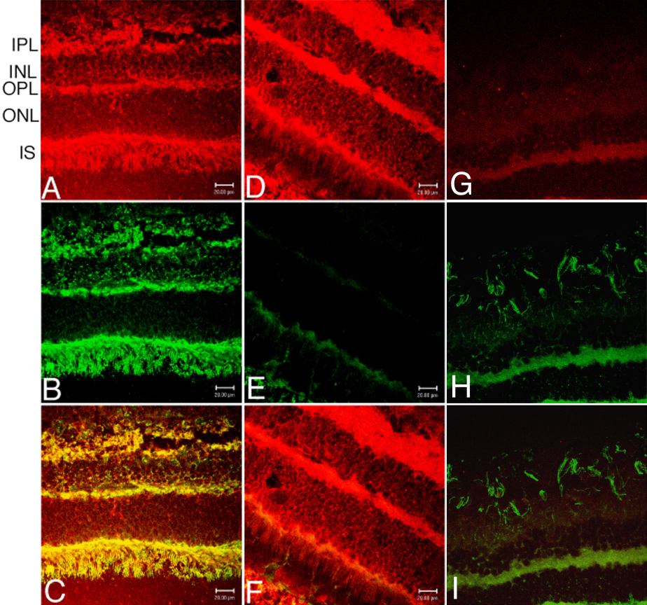
Figure 3. Confocal microscopy of the primte retina
Confocal microscopy of the retina from the gene-injected monkey (A-C, G-I) and from the control, un-injected monkey (D-F). Immunoreactive insulin receptor, stained with rhodamine-labeled secondary antibody, is shown in Panels A and D. Immunoreactive β-galactosidase, stained with the fluorescein-labeled secondary antibody, is shown in Panels B and E. Staining with the pre-immune mouse IgG or the rabbit IgG is shown in Panels G and H. The respective superimposed images are shown in panels C, F, and I. The yellow image in panel C shows co-localization of the β-galactosidase and human insulin receptor in the retina of the gene-injected monkey, including the photoreceptor cells. Scale bar: 20 μm. Abbreviations in Panel A: IPL, inner plexiform layer; INL, inner nuclear layer; OPL, outer plexiform layer; ONL, outer nuclear layer; IS, inner segments. All specimens shown in Panels A-C were taken 48 h after the intravenous administration of clone 756, the pSV40-β-galactosidase plasmid in monkey 1 (see Methods).