![]() Figure 2 of
Zhang, Mol Vis 2003;
9:465-472.
Figure 2 of
Zhang, Mol Vis 2003;
9:465-472.
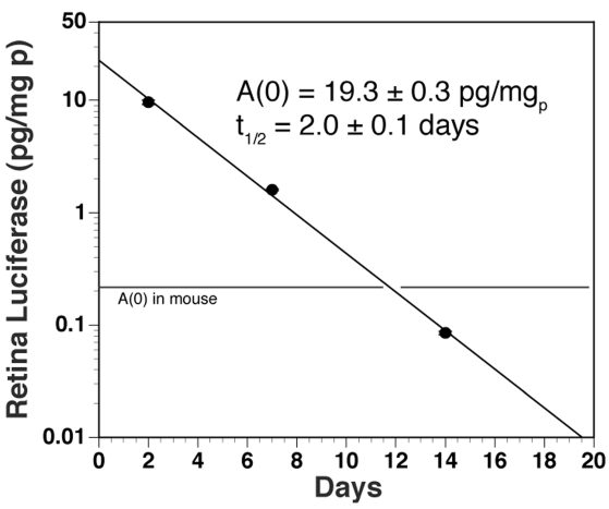
Figure 2. Luciferase gene expression in the primate eye
Retinal luciferase activity in 3 rhesus monkeys sacrificed at 2, 7, or 14 days after intravenous administration of clone 790 in monkeys 1, 2, and 3, respectively (see Methods). The data were fit to a single-exponential equation to yield the slope/half-time (t1/2) and the intercept, [A(0)]. The horizontal line shows the A(0) of luciferase activity in control mouse brain, 0.22±0.08 pg/mg [20], which indicates the level of gene expression in the monkey retina is still above the therapeutic level for at least 2 weeks after injection. The luciferase expression plasmid was under the influence of the SV40 promoter in these studies. The diagonal line through the data points was drawn by eye. Error bars representing the stardard deviation are shown over each closed circle.