![]() Figure 1 of
Zhang, Mol Vis 2003;
9:465-472.
Figure 1 of
Zhang, Mol Vis 2003;
9:465-472.
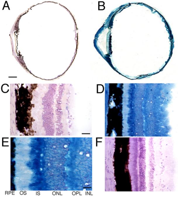
Figure 1. β-Galactosidase histochemistry of primate eyes
β-galactosidase histochemistry in control, un-injected Rhesus monkey eye (Panels A and C) and gene-injected Rhesus monkey eye (Panels B, D, E, and F). Panels A-E show β-galactosidase histochemistry followed by counter-staining with hematoxylin, whereas Panel F shows the retina only counter-stained with hematoxylin. Magnification in Panels A and B is the same; the magnification bar in Panel A represents 2 mm. The magnification in Panels C, D, and F is the same; the magnification bar in Panel C represents 32 μm. The magnification bar in Panel E represents 13 μm. Abbreviations in Panel E: INL, inner nuclear layer; OPL, outer plexiform layer; ONL, outer nuclear layer; IS inner segments; OS, outer segments; RPE, retinal pigmented epithelium. All specimens shown in Panels B, D-F were taken 48 h after the intravenous administration of clone 756, the pSV40-β-galactosidase plasmid, in monkey 1 (see Methods).