![]() Figure 5 of
Morin, Mol Vis 2003;
9:449-459.
Figure 5 of
Morin, Mol Vis 2003;
9:449-459.
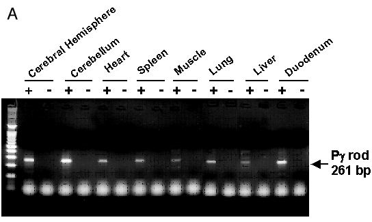
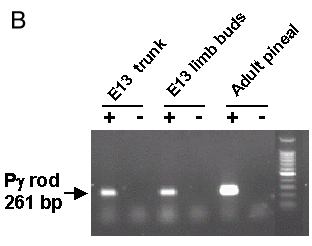
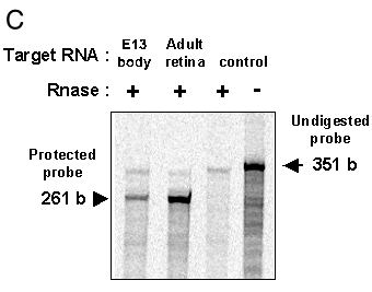
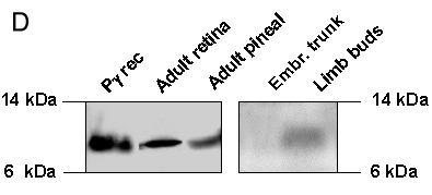
Figure 5. RT-PCR, RPA and western blot analyses of Pγ-rod expression in adult and embryonic rat tissues
A: Total RNA (3 μg) from the indicated adult rat tissues was reverse transcribed, followed by 35 cycles PCR amplifications with Pγ-rod primers. Test reactions (+) were run in parallel with control reactions lacking reverse transcriptase (-). The reaction products (1/5 of the reaction) were analyzed by electrophoresis and visualized under UV light. Standard: 100 bp ladder (Promega). B: Total RNA (3 μg from trunk or limb buds of E13 rat embryos or 0.3 μg from adult rat pineal) was reverse transcribed, followed by 30 cycles PCR amplification with Pγ-rod primers. Test reactions (+), controls without reverse transcriptase (-). The reaction products (1/5 of the reaction) were analyzed as above. C: Aliquots of target RNA from the indicated tissues were hybridized with the Pγ-rod 35S-riboprobe and subjected to digestion with RNase A/T1. Samples were electrophoresed through 6% acrylamide / 6 M urea gel. The gel was dried and exposed 3 days to a PhosphoImager screen. Lane 1: polyA+ RNA (15 μg) from rat embryonic body (trunk+limb buds). Lane 2: total RNA (1 μg) from adult rat retina. Lane 3: yeast total RNA (50 μg). Lane 4: yeast total RNA (50 μg) without RNase. D: Protein extracts prepared as described in Experimental Procedures, were analyzed by SDS-PAGE and western blotted (retina 20 μg, pineal 20 μg, embryonic trunk 20 μg, limb buds 20 μg, recombinant Pγ 50 ng). The membranes were incubated with an antibody directed against the c-terminal region of Pγ-rod (1:15,000) and with an antibody directed against its N-terminal region (1:500). Immunoreactions were developped with peroxydase-coupled anti-rabbit (1:3000) and the enhanced chemiluminescence detection kit (Amersham).