![]() Figure 4 of
Morin, Mol Vis 2003;
9:449-459.
Figure 4 of
Morin, Mol Vis 2003;
9:449-459.
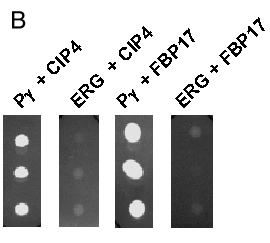
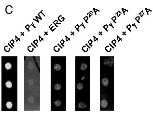
Figure 4. Interaction of Pγ-rod with CIP4, another SH3-containing protein
A: alignment of the SH3 domains of CIP4 and FBP17: identical amino acids are indicated by (:). B: L40 yeast expressing the Pγ-rod bait (as lexADBD fusion) were transformed with either CIP4 or FBP17 (as gal4AD fusions) and plated as triplicate drops on medium lacking histidine. Negative controls were performed in parallel using L40 yeast expressing the neutral bait ERG19 (ERG). C: The indicated proline mutants of Pγ-rod (as lexADB fusions) were transformed into L40 yeast expressing CIP4 (as gal4AD fusion) and plated as triplicate drops on medium lacking histidine. Positive control corresponds to wild-type (WT) Pγ-rod. Negative control corresponds to the neutral bait ERG19 (ERG).
A:
CIP4:  SPIGQCVAIY HFEGSSEGTV SMSEGEDLSL MEEDKGDGWT RVRRKQGGEG YVPTSYLRVT LN
         :: : : :  :::  :::  :  ::: ::   ::::::::: : ::    :: ::::::  :  :
FBP17: PAIGTCKALY TFEGQNEGTI SVVEGETLSV IEEDKGDGWT RIRRNEDEEG YVPTSYVEVY LD
|