![]() Figure 2 of
Morin, Mol Vis 2003;
9:449-459.
Figure 2 of
Morin, Mol Vis 2003;
9:449-459.
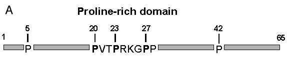
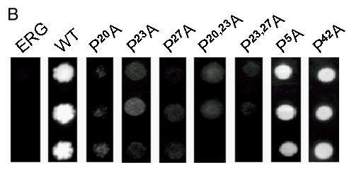
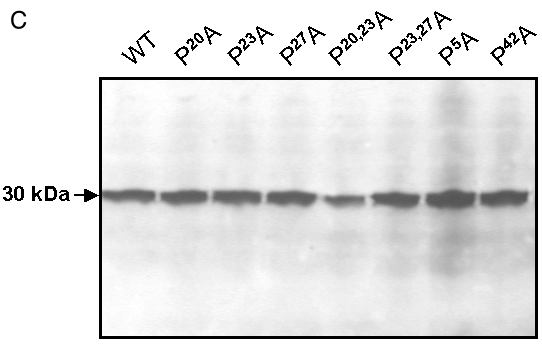
Figure 2. Mutations of the Pγ-rod proline-rich domain interrupt binding to FBP17
A: Scheme of the proline mutations within the Pγ-rod bait. B: The indicated proline mutants of Pγ-rod (as lexADBD fusions) were transformed into L40 yeast expressing FBP17 (as gal4AD fusion) and plated as triplicate drops on medium lacking histidine. Identical results were obtained with 3 to 5 independent clones of each mutant. Positive control corresponds to wild-type (WT) Pγ-rod. Negative control corresponds to the neutral bait ERG19 (ERG). C: Protein extracts from the cotransformants described above were analyzed by western blot to verify that wild type and mutated forms of Pγ-rod were correctly expressed. The lexADBD-Pγ-rod fusion proteins were detected with anti-lexA antibody as described in "Experimental procedures".