![]() Figure 5 of
Clout, Mol Vis 2003;
9:440-448.
Figure 5 of
Clout, Mol Vis 2003;
9:440-448.
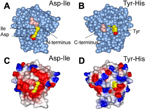
Figure 5. Residues implicated in binding integrin viewed on the surface of domain 4 of βig-h3
A and B: Space-filling representation of domain 4 of βig-h3. The two views are related by a 180° rotation about a vertical axis. The N- and C-termini of the domain are colored pink and labelled (Met502 and Pro634 respectively). The postulated integrin binding amino acids are in yellow (carbon atoms) and red (oxygen atoms) and are labelled. C and D: Electrostatic surface representations in identical orientations to A and B, respectively. Positive and negative potential is indicated by blue and red coloring, respectively. Residues implicated in integrin binding are colored yellow and red as in A and B.