![]() Figure 2 of
Clout, Mol Vis 2003;
9:440-448.
Figure 2 of
Clout, Mol Vis 2003;
9:440-448.
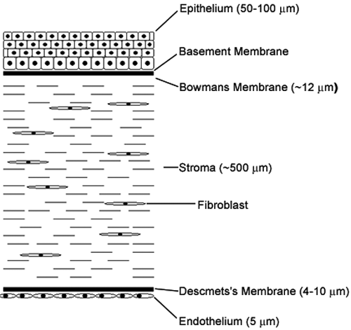
Figure 2. Schematic transverse section through the cornea
In normal corneas the βig-h3 protein is detected throughout the stroma, in Bowman's layer and in endothelial cells [6,8,11-13]. In healing and diseased corneas, βig-h3 has been identified in the retrocorneal fibrous membrane (which develops posterior to the pre-existing Descemet's membrane) in the stromal keratocytes [13,14], and as deposits in the corneal stroma in the case of corneal dystrophies [12].