![]() Figure 1 of
Clout, Mol Vis 2003;
9:440-448.
Figure 1 of
Clout, Mol Vis 2003;
9:440-448.
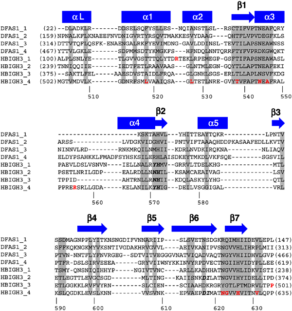
Figure 1. Sequence alignment of selected FAS1 domains
The sequences are of Drosophila melanogaster fasciclin I (P10674) and human βig-h3 (Q15582). The four domains of each protein are included in the alignment and the sequence numbers are indicated in brackets at the start and end of each FAS1 domain. Additional numbering is provided, below the alignment, for the fourth FAS1 domain of βig-h3. The secondary structure elements are indicated above the alignment. Conserved residues are shaded in grey. βig-h3 residues mutated in corneal dystrophies are in red, and residues implicated in integrin binding to βig-h3 are in bold and italic.