![]() Figure 5 of
Xi, Mol Vis 2003;
9:410-419.
Figure 5 of
Xi, Mol Vis 2003;
9:410-419.
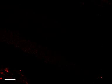
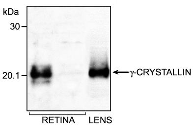
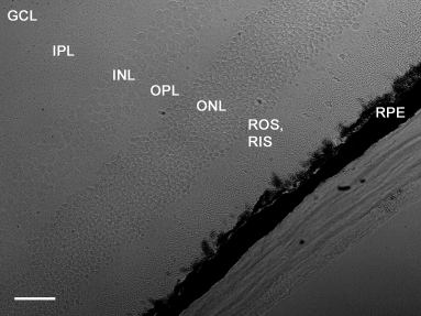
Figure 5. Expression of γ-crystallin in mouse retinas
Expression of γ-crystallin in adult mouse retinas by immunoblotting and immunofluorescence. Immunoblot analysis. (A) Cell lysates were prepared from 12.5 week old retinas and analyzed by SDS-PAGE and immunoblotting. A polyclonal antibody against bovine γ-crystallin fraction was used. Lanes are: left and middle, mouse retina; right, bovine lens γ-crystallin (0.7 μg). Note the significant variability of expression of γ-crystallin in retina derived from two animals of the same litter. Immunofluorescence (B) γ-crystallin (red) was localized using a polyclonal antibody against bovine γ-crystallin fraction. Note that γ-crystallin was distributed in the ganglion cell layer, inner photoreceptor layer, and outer nuclear layer. Cellular morphology was visualized with differential interference contrast. A DIC image of a normal retina is shown in (C). Pre-adsorption of the primary antibody with bovine γ-crystallin protein (negative control) showed no detectable γ-crystallin immunofluorescence (D) in the retina shown in (C). Bar represents 13 μm (B-D).
A:
B:
C:
D: