![]() Figure 2 of
Xi, Mol Vis 2003;
9:410-419.
Figure 2 of
Xi, Mol Vis 2003;
9:410-419.
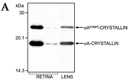
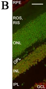
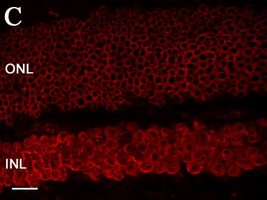
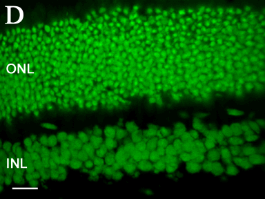
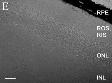
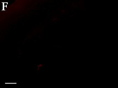
Figure 2. Expression of αA-crystallin in mouse retinas
Expression of αA-crystallin in adult mouse retinas by immunoblotting and immunofluorescence. Immunoblot analysis. (A) Cell lysates were prepared from 12.5 week old retinas and analyzed by SDS-PAGE and immunoblotting. A 20 μg aliquot of the retinal protein was applied to the gel. The monoclonal antibody against bovine αA-crystallin was used. Lanes are: left and middle, mouse retinas of two different animals; right, mouse lens epithelial cells (5x10⁴, corresponding to 0.3 μg of αA-crystallin). Note the significant variability in expression of αA-crystallin in retina derived from two animals of the same litter. Note also that the αA- and αA-insert proteins from the retinas had the same mobility as the proteins from lens epithelial cells. Immunofluorescence. (B) αA-crystallin (red) was localized using an antibody to αA-crystallin, and nuclei (green) were stained with TOTO-1. Cellular morphology was visualized with differential interference contrast (DIC). Note that αA-crystallin was distributed in the ganglion cell layer, inner photoreceptor layer, and outer nuclear layer (B). A higher magnification of αA-crystallin distribution in the inner and outer nuclear layers of the retina is shown in (C). Note that αA-crystallin distribution was restricted to the membranes of the outer nuclear layers, but it is also distributed in the structures within the nucleus of the inner nuclear layer. A high magnification image of TOTO-1 immunofluorescence of the nuclei shown in (C) is shown in (D). A DIC image of an αA-/- retina (negative control) is shown in (E). No αA-crystallin immunofluorescence was detectable in the αA-/- mouse retina. Bar represents 13 μm (B, E, F); 5 μm (C,D). RPE, retinal pigment epithelium; ROS, rod outer segments; RIS, rod inner segments; ONL, outer nuclear layer; OPL, outer plexiform layer; INL, inner nuclear layer; IPL, inner plexiform layer; GCL, ganglion cell layer.