![]() Figure 7 of
O'Brien, Mol Vis 2003;
9:401-409.
Figure 7 of
O'Brien, Mol Vis 2003;
9:401-409.
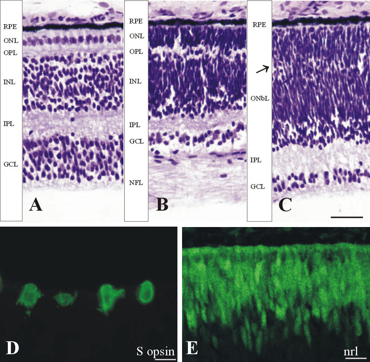
Figure 7. Human retina at fetal weeks 18-19
Cresyl violet stained frozen sections from a retina at Fwk 18 (A,B,C). All layers in the fovea (A) and optic disc (B) are more mature. The ONL at the optic disc is much thicker due to the addition of rods (C). In the periphery, a thick IPL is now present and a thin discontinuous OPL (arrow) is dividing the OnbL into ONL and INL. D,E: ICC labeled S cones (D) and Nrl-labeled rod nuclei (E) are present near the retina edge in the outer retina, despite its immature appearance. The scale bars represent 25 μm in A through C, 12 μm in D, and 8 μm in E.