![]() Figure 5 of
O'Brien, Mol Vis 2003;
9:401-409.
Figure 5 of
O'Brien, Mol Vis 2003;
9:401-409.
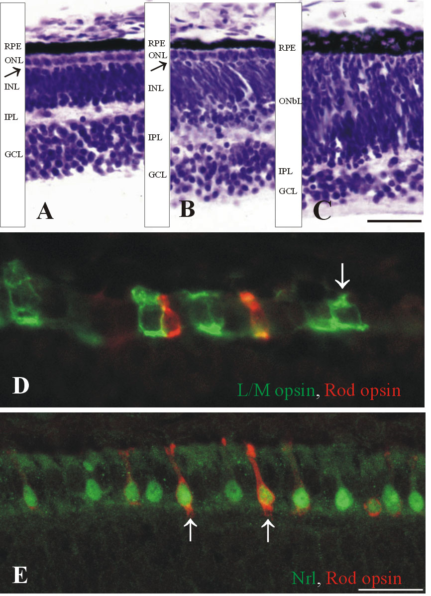
Figure 5. Human retina at Fwk 14-16
A,B,C: Cresyl violet stained frozen sections from a Fwk 14 retina. The foveal region (A) has more mature cones in the ONL and the OPL (arrow) is more apparent. B: At the eccentricity of the disc, a thin ONL and think INL are present with a thin OPL separating them (arrow). C: In the peripheral nasal retina, a thin IPL separates the GCL from a thick OnbL. D: A section on the edge of the Fwk 16 fovea is double labeled for L/M opsin (green) and Rod opsin (red). The arrow indicates a heavily labeled cone outer segment. E: Section on the foveal edge at Fwk 16 double labeled for Nrl (green) and Rod opsin (red). All rods have a Nrl-labeled nucleus (green), but only a few express Rod opsin (arrows). The scale bars represent 25 μm in A through C, and 12 μm in D and E.