![]() Figure 4 of
O'Brien, Mol Vis 2003;
9:401-409.
Figure 4 of
O'Brien, Mol Vis 2003;
9:401-409.
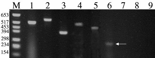
Figure 4. Expression of actin, Crx, PDEB, IRBP, TULP, and Nrl in the Fwk 12 human eye
An RT-PCR agarose gel stained with ethidium brominde from a Fwk 12 human retina. Bands were present in Lanes 1-3 corresponding to actin, Crx, PDEB, and were detected for the first time for IRBP (lane 4), TULP (lane 5) and Nrl (lane 6). M indicates the DNA molecular weight marker. Lanes 7 (S opsin), 8 (L/M opsin), and 9 (Rod opsin) have no detectable bands.