![]() Figure 3 of
O'Brien, Mol Vis 2003;
9:401-409.
Figure 3 of
O'Brien, Mol Vis 2003;
9:401-409.
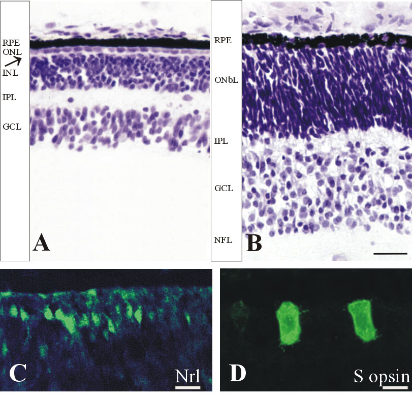
Figure 3. Human retina at Fwk 11-14
A,B: Cresyl violet stained sections from a Fwk 12 retina showing the fovea (A) and the retina near the optic disc (B). Foveal morphology (A) has changed relatively little except that the OPL is more apparent (arrow). At the optic disc (B), a thin IPL now separates off the GCL and a nerve fiber layer (NFL) is present. C: Nrl-positive nuclei (green) present at the peripheral edge of Nrl expression, which is 3.6 mm from the fovea, at Fwk 13. These nuclei form a row under the unstained cones. D: S opsin (green) is detected in a subset of cones across the entire central retina by Fwk 14. The scale bars represent 25 μm in A and B, 15 μm in C, and 10 μm in D.