![]() Figure 2 of
O'Brien, Mol Vis 2003;
9:401-409.
Figure 2 of
O'Brien, Mol Vis 2003;
9:401-409.
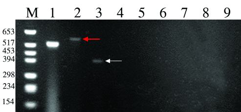
Figure 2. Expression of actin, Crx, and PDEB in the Fwk 10 human eye
A reverse transcription polymerase chain reaction (RT-PCR) agarose gel stained with ethidium brominde from a Fwk 10 human retina. Bands were only detected in lanes 1-3 which correspond to actin, Crx (red arrow) and PDEB (white arrow), respectively. M indicates the DNA molecular weight marker. 1, actin; 2, Crx; 3, PDEB; 4, IRBP; 5, TULP; 6, Nrl; 7, S opsin; 8, L/M opsin and 9, Rod opsin.