![]() Figure 1 of
O'Brien, Mol Vis 2003;
9:401-409.
Figure 1 of
O'Brien, Mol Vis 2003;
9:401-409.
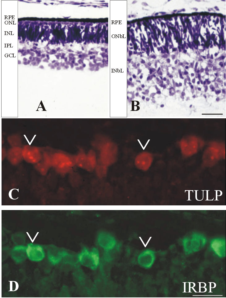
Figure 1. Human retina at Fwk 9-10
A,B: Cresyl violet stained frozen sections of a Fwk 10 retina showing the fovea (A) and the retina near the optic disc (B). The fovea has a single thin layer of cone photoreceptors forming the outer nuclear (ONL) adjacent to the pigment retina pigment epithelium (RPE). An immature inner nuclear layer (INL) is separated from the ganglion cell layer (GCL) by a thin inner plexiform layer (INL). B: Retina near the optic disc is thicker and is divided into the outer neuroblastic layer (ONbL) and inner neuroblastic layer (INbL). C,D: The same section from the Fwk 9 fovea showing immunocytochemical labeling for TULP (C) and IRBP (D). Most of the cones are double labeled (arrowheads). The scale bars represent 25 μm in A and B; in C and D they represent 10 μm.