![]() Figure 4 of
Girao, Mol Vis 2003;
9:24-30.
Figure 4 of
Girao, Mol Vis 2003;
9:24-30.
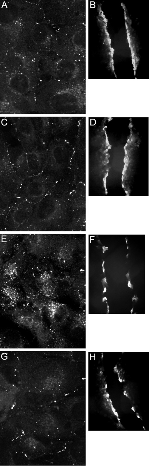
Figure 4. Effect of phosphorylation and proteasome inhibitors on intercellular communication and subcellular distribution of Cx43
LEC were either treated with 40 μM MG132 (C and D) for 1 h or 50 ng/ml TPA (E and F) for 30 min; simultaneous treatment with MG132 and TPA was performed by incubation with 40 μM MG132, for 1 h, and 50 ng/ml TPA was added for the last 30 min (G and H). Cells incubated in the absence of TPA and MG132 were used as controls (A and B). The cells were then either assayed for intercellular communication by Lucifer yellow dye transfer, after scrape loading (B, D, F, and H, magnification 200x), or fixed and stained with antibodies directed against Cx43 and imaged by confocal microscopy (A, C, E, and G). Intercellular communication was evaluated as the average distance traveled by the dye Lucifer yellow along the monolayer and is represented as a histogram (I). The values are the average of five individual experiments with a coefficient of variation typically inferior to 6%.