![]() Figure 1 of
Girao, Mol Vis 2003;
9:24-30.
Figure 1 of
Girao, Mol Vis 2003;
9:24-30.
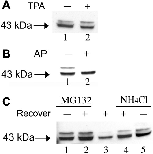
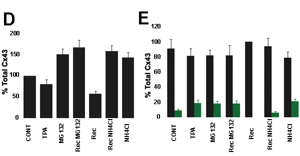
Figure 1. Immunodetection of Cx43 in lens epithelial cells
Immunodetection of Cx43 in lens epithelial cells treated with TPA, in the presence of lysosome or proteasome inhibitors. A: Cell lysates of LEC exposed to 50 ng/ml TPA for 30 min were western blotted and probed with monoclonal antibodies directed against Cx43 (lane 2). Control cells were incubated in the absence of TPA (lane 1). B: Enzymatic dephosphorylation of Cx43 from lens epithelial cells previously incubated with TPA. Cell lysates were incubated with alkaline phosphatase from E. coli at 37 °C overnight (lane 2). Controls were incubated in the absence of alkaline phosphatase (lane 1). C: LEC were exposed to 50 ng/ml TPA for 30 min, in the presence of 40 μM MG132 (lanes 1 and 2) or 10 mM NH4Cl (lanes 4 and 5). Following treatment with 50 ng/ml TPA for 30 min, the cells recovered either in the absence (lane 3) or presence of 40 μM MG132 (lane 2) or 10 mM NH4Cl (lane 4) during 2 h. Proteins in cell lysates were separated by SDS-PAGE, transferred to PVDF membranes and probed with antibodies to Cx43. D: Changes in the amount of total Cx43 in epithelial cells, as determined by densitometric analysis of the films. Values are expressed as percentage of non-treated controls. E: Quantification of the relative amount of phosphorylated (green) and non-phosphorylated (black) forms of Cx43. Values are expressed as percentage of the total Cx43 present in each lane. Data is the average (with standard deviation) of five (A) or three (C) independent experiments.