![]() Figure 1 of
Hedegaard, Mol Vis 2003;
9:355-359.
Figure 1 of
Hedegaard, Mol Vis 2003;
9:355-359.
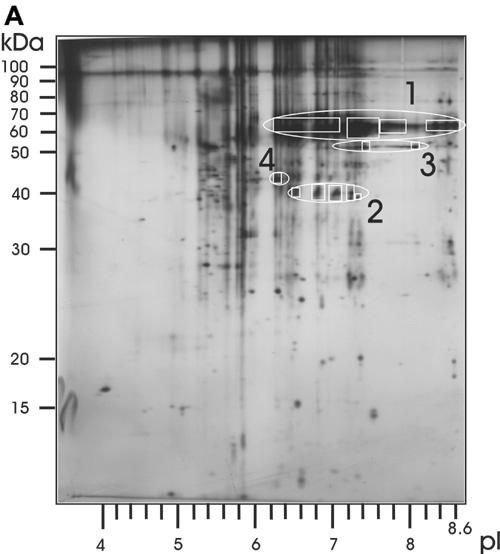
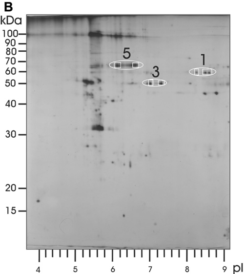
Figure 1. Two-dimensional gel electrophoresis
Two-dimensional gel electrophoresis performed in 1987 on proteins extracted from one cornea with granular corneal dystrophy type III (Reis-Bücklers, A) and one normal human cornea (B). Proteins were run in the first dimension from pH 3.5 to pH 10 and in the second dimension in a 12.5% acrylamide gel. In 2002, the proteins within the encircled areas were identified by mass spectrometry. Area 1 was identified as TGFBIp (63 kDa), area 2 was TGFBIp fragments (40 kDa), area 3 was aldehyde dehydrogenase class 3 (50 kDa), area 4 was actin (42 kDa), and area 5 was albumin (66 kDa). Note that the high amount of TGFBIp obscures detection of albumin in the cornea with GCDIII. Boxes mark the areas where gel plugs were excised and analyzed.