![]() Figure 1 of
Chen, Mol Vis 2003;
9:345-354.
Figure 1 of
Chen, Mol Vis 2003;
9:345-354.
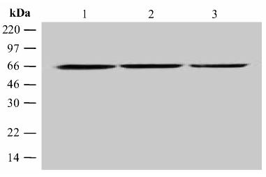
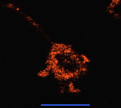
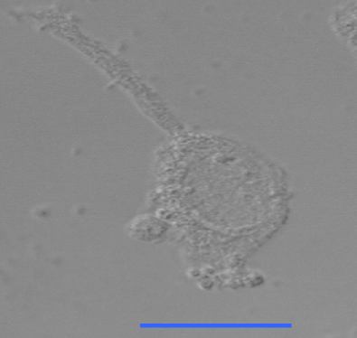
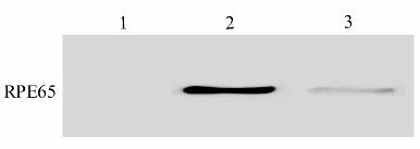
Figure 1. Subcellular localization of RPE65 in HEK293 cells
A: Confocal microscope image with a Rhodamine filter to demonstrate the localization of RPE65 within the HEK293 cell. Bars correspond to 50 μm. B: Bright field image of the same cell. Bars correspond to 50 μm. C: Western blot analysis of the cell fractions after rate-zonal centrifugation. Lane 1 represents the nuclear fraction; lane 2, the microsomal fraction; and Lane 3, the soluble fraction. D: Analysis of RPE65 antibody specificity. Western blot analysis was performed on HEK293 cells with purified recombinant human RPE65 [37] and bovine RPE homogenate (as control). The primary antibody was anti-RPE65 polyclonal antibody (1:1,000 dilution). Lane 1, purified recombinant human RPE65 protein (1 μg); Lane 2, bovine RPE crude preparation (5 μg); and Lane 3, HEK293 cell extract (100 μg) prepared as described in Methods.
A:
B:
C:
D: