![]() Figure 1 of
Kaldi, Mol Vis 2003;
9:337-344.
Figure 1 of
Kaldi, Mol Vis 2003;
9:337-344.
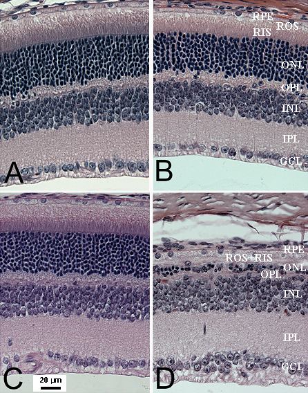
Figure 1. Light micrographs of mouse retinas
Sections were taken about 1 mm from the optic nerve head along the superior meridian. A: Bright-reared, control. B: Bright-reared, light-stressed; C: Dim-reared, control. D: Dim-reared, light-stressed. Abbreviations: RPE, retinal pigment epithelium; ROS, rod outer segments; RIS, rod inner segments; ONL, outer nuclear layer; OPL, outer plexiform layer; INL, inner nuclear layer; IPL, inner plexiform layer; and GCL, ganglion cell layer. (H & E staining)